Spine Treatment
What I do
Spin treatment
Spinal Pain
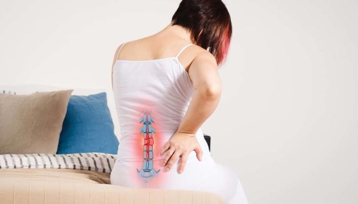
Spinal pain in the lumbar region (lower back) and cervical region (neck) are highly prevalent and are often the causes for many lost work days. Lumbar muscle strains and sprains are the most common causes of low back pain. The thoracic spine can also be a site of spinal pain, but because it is much more rigid, the thoracic spinal area is much less frequently injured than the lumbar and cervical spine.
The lumbar and cervical spine are prone to strain because of its weight-bearing function and involvement in moving, twisting and bending. Lumbar muscle strain is caused when muscle fibers are abnormally stretched or torn. Lumbar sprain is caused when ligaments — the tough bands of tissue that hold bones together — are unusually stretched. Both of these can result from a sudden injury or from gradual overuse.
Symptoms
Non-surgical low back, cervical and thoracic pain usually affects the central or para-spinal soft tissue without radiating into the arms, around the chest or down the legs. On the contrary, pain radiating from the spine into the extremities or chest wall implies structural pinching of the nerves in the spine that might require a surgical opinion if the situation fails to improve within days to weeks with non-surgical symptomatic treatment.
Other symptoms include:
- Stiffness in the low back area, restricting range of motion
- Inability to maintain normal posture due to stiffness and/or pain
- Muscle spasms either with activity or at rest
- Pain that persists for a maximum of 10-14 days
- Notable loss of motor function such as the ability to tiptoe or heel walk.
Diagnosis
Imaging tests
These tests may include:
- X-rays. An X-ray of your back can reveal bony changes, such as bone spurs that may be narrowing the space within the spinal canal. Each X-ray involves a small exposure to radiation.
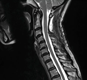
- Magnetic resonance imaging (MRI). An MRI uses a powerful magnet and radio waves to produce cross-sectional images of your spine. The test can detect damage to your disks and ligaments, as well as the presence of tumors. Most important, it can show where the nerves in the spinal cord are being pressured.
- CT or CT myelogram. If you can’t have an MRI, your doctor may recommend computerized tomography (CT), a test that combines X-ray images taken from many different angles to produce detailed, cross-sectional images of your body. In a CT myelogram, the CT scan is conducted after a contrast dye is injected. The dye outlines the spinal cord and nerves, and it can reveal herniated disks, bone spurs and tumors.
Physical Therapy for Spinal Stenosis
A physical therapy program can go a long way toward easing your symptoms and can also help with:
- Flexibility
- Balance
- Endurance
Spinal stenosis exercises
Talk with your doctor or physical therapist about exercises that can help you:
- Gain flexibility: Stretching exercises can help with pain and make it easier to hold and move your neck and spine in healthier ways.
- Strengthen your muscles: A series of exercises called stabilization training can help build up the muscles that support your neck and give them better balance. Like stretching, these are simple exercises you can do at home without any special equipment.
- Boost your fitness: Aerobic exercises, ones that get your heart and breathing rates up, release chemicals called endorphins that can ease pain. Examples of aerobic exercise include bicycling or swimming.